Estimated to account for ~2-3% of all ED visits per year, dizziness is a complaint that we have all dealt with as ED doctors. Despite being so common, working up dizziness can be quite confusing as there are many different components to consider, from the exam to the history, special tests and imaging. Thankfully, there has recently been more consensus on what the most important factors are to narrow our differential and minimize missing dangerous diagnoses.
Breaking down the dizziness workup into 3 steps has helped me to standardize and simplify a complex complaint, but before that I’d like to discuss some key points to focus on within those 3 steps, as well as stress the utility of learning the HINTS exam and Dix-Hallpike maneuvers.
First Things First: Is something else going on?

Many patients will present with dizziness and something else (chest pain, difficulty breathing, vomiting, to name a few). Before going down the wormhole that is dizziness, make sure to consider all the other things that can make a patient feel “unsteady, like the room is spinning, like I’m going to pass out” (more on these later). Dizziness is one of the ED complaints in which a thorough review of systems will truly change your workup. Run your usual list of life-threats for each body system in your head to make sure that your posterior stroke patient isn’t actually septic, pregnant, or having melenic stool.
Acute vs. Episodic, THEN Central vs. Peripheral
The biggest paradigm shift in the evaluation of dizziness in the ED is the concept of Acute Vestibular syndrome (AVS) and Episodic Vestibular Syndrome (EVS). Although the distinction between a central and peripheral cause of vertigo is important, delineating between AVS and EVS first is more helpful in interpreting your physical exam, focusing your workup, and narrowing your differential. AVS and EVS are typically defined as the following:
Acute Vestibular Syndrome
Acute-onset, constant dizziness lasting days-weeks with vestibular symptoms (vomiting, nystagmus, ataxia) even at rest. Typically is worse with head/body movement. Differential diagnoses of AVS include:
- Vestibular neuritis
- Labyrinthitis
- Stroke
Episodic Vestibular Syndrome
Transient dizziness with short-lived vestibular symptoms that completely resolve at rest. EVS is sometimes further subdivided based on triggers and duration of symptoms into spontaneous EVS (s-EVS) and transient EVS (t-EVS).
s-EVS does not have a clear trigger but can be worse with head/body movement & typically lasts minutes-hours. Differential includes:
- Meniere Disease
- Vestibular Migraine
- TIA
t-EVS does have clear triggers, usually head movement & typically lasts seconds-minutes. Differential includes:
- Orthostasis
- BPPV
The T’s and The D’s
I attempt to simplify the most important parts of the history and physical exam of a dizzy patient as the “2 T’s” and “5 D’s”
The “2 T’s”: Timing and Triggers
Getting a thorough idea of the timing of your patient’s dizziness as well as triggers (if they exist) will stratify them into one of the above categories of AVS, s-EVS and t-EVS.
The “5 D’s”: Do not pass go; Go directly to MRI
During your history and clinical exam, if any of these 5 D’s are present in conjunction with dizziness, you should strongly consider sending your patient for advanced imaging (specifically MRI) as these are all associated with cerebellar pathology from a posterior circulation stroke:
- Diplopia (double-vision), concerning for a CN VI, IV, or III lesion
- Dysarthria (”marbles in the mouth”), concerning for a CN VII lesion
- Dysphagia (difficulty swallowing or voice change), concerning for a CN IX or X lesion
- Dysmetria (gait, tandem gait, finger-to-nose, Romberg), concerning for cerebellar damage
- Dysdiadochokinesia (inability to perform rapid alternating movements), concerning for cerebellar damage
Sidenote: The HINTS Exam
The HINTS (Head Impulse, Nystagmus, Test of Skew) exam is often thought of as a tool that we can use in the ED to “rule out a stroke” and make the diagnosis of BPPV, when this is not the case. The HINTS exam is only used in cases of AVS, it is not used in cases of EVS, in which BPPV is a differential. The HINTS exam is useful if trying to differentiate between a central and peripheral cause of AVS (for example, vestibular neuritis vs a stroke) but should not be used to rule out the diagnosis of stroke in someone who is intermittently dizzy. I’ll touch more on this later.
Now with these important points discussed, let’s work through the 3 step approach to dizziness:
The 3-Step Approach to Dizziness
1. The Story
Take time to obtain a thorough history, paying special attention to the timing and triggers as discussed above. Important questions to ask are when it started, is it constant or intermittent, what makes it worse, and how long does it last when it onsets. Make sure to run a good review of systems to make sure you’re not dealing with an entirely different workup.
2. The Exam
Every dizzy patient deserves a full neurologic exam, including extraocular movement, cranial nerve and cerebellar exams. I won’t recite the entire neuro exam in full, but here are some points to focus on in the dizzy patient:
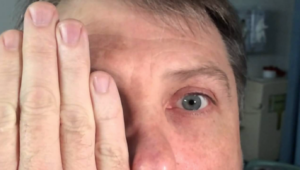
- Uni-directional, fatigable nystagmus is the only reassuring type of nystagmus that could potentially be a benign cause, such as BPPV. Bi-directional, non-fatigable, vertical, and rotary nystagmus are all concerning.
- Nystagmus is a normal finding if you make your patient look way off the midline. To assess for nystagmus, move your focal point only about 20-30 degrees from the midline.
- Don’t forget to look in the ears, especially if they have any audiologic symptoms (ear ringing, ear pain); you can catch anything from a cerumen impaction to a developing Ramsay-Hunt syndrome.
- While it may be difficult to assess a patient’s gait as we don’t know their baseline, keep in mind that nearly all posterior stroke patients will have an abnormal gait assessment, especially with tandem gait.
- Remember the 5 D’s. If any are present, go straight to MRI.
3. The MRI
The hard work is done, now it’s time to make a decision: does your patient need an MRI?
Put simply, if your patient:
- Sounds like EVS, but you cannot confirm the diagnosis with a Dix-Hallpike maneuver
- Sound like AVS, but you cannot reliably rule out a central cause with the HINTS exam
- Has any focal neurologic deficit, including any cerebellar or cranial nerve deficit
- Is unable to ambulate with a steady gait
- Has any type of nystagmus that is not unidirectional and horizontal
- Is elderly and has stroke risk factors
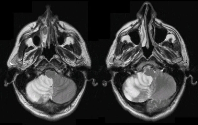
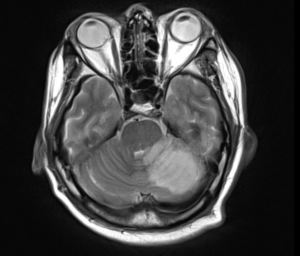
You need an MRI to assess for posterior circulation stroke. Be aware that even MRI has an overall sensitivity of ~80-90% in early (<48h) posterior stroke.
The Ultimate Goal: Learning the HINTS and Dix-Hallpike

In our busy EDs, especially in the era of waiting room medicine, many of us are hesitant to take the time to do an additional exam on our dizzy patients who are already getting a very detailed history and physical exam. However, as evidenced above, I argue that being able to perform the HINTS exam and Dix-Hallpike maneuver correctly and reliably is a huge skill we can use to help our dizzy patients, as we may be able to save them from a very slow and very expensive test (MRI) by just using our hands.
For example,
Is this a dizziness that comes and goes, lasts anywhere from seconds to minutes or hours, is worse with body and head movement but resolves with rest? Sound like Episodic Vestibular Syndrome (EVS).
- If you can confirm the diagnosis of BPPV with a Dix-Hallpike maneuver, then you do not need an MRI.
Is this a dizziness that has been going on for days, always seems to be there, does get worse but never goes away? Sounds like Acute Vestibular Syndrome (AVS).
- If you can confirm the diagnosis of vestibular neuritis with a HINTS exam, then you do not need an MRI.
Literature has shown that ED providers have a long way to go in mastering these exams, specifically how to correctly perform them and in which cases to properly implement them. On the other hand, literature has also shown that once ED providers are trained in its use, the HINTS exam can be up to 97% sensitive for stroke. There is much to gain and even more time and money to save by helping our patients avoid an MRI.
Conclusion
It seems like the deeper one dives into the topic of dizziness, the more complicated and confusing it becomes. Having a simple step-by-step approach for this common complaint, as well as an understanding of the bedside tests and most important historical factors, can help us to efficiently and confidently work up these patients in the ED.
References
- The majority of this post was sourced by the SAEM GRACE-3 paper on dizziness and vertigo in the ED:
- Edlow JA, Carpenter C, Akhter M, Khoujah D, Marcolini E, Meurer WJ, Morrill D, Naples JG, Ohle R, Omron R, Sharif S, Siket M, Upadhye S, E Silva LOJ, Sundberg E, Tartt K, Vanni S, Newman-Toker DE, Bellolio F. Guidelines for reasonable and appropriate care in the emergency department 3 (GRACE-3): Acute dizziness and vertigo in the emergency department. Acad Emerg Med. 2023 May;30(5):442-486. doi: 10.1111/acem.14728. PMID: 37166022.
- EMRAP C3: “Management of Dizziness”:
- Stuart Swadron MD, Jessica Mason MD, Mel Herbert MD (Hosts). (2017, April). Management of Dizziness [Audio Podcast Episode]. In Emergency Medicine Research and Perspectives. EMRAP. https://www.emrap.org/episode/c3dizziness/c3dizziness2
- Shoutout to recent Copa alumnus Dr. Jonathan Kelley, who gave an excellent presentation on this topic during our resident conference. Many of the important points from this post came from my notes on his presentation. Thanks Jon!